InformationPrimary endodontic treatment Endodontic retreatment Special treatments Endodontics step by stepProduct catalogueOrder and payment information Previous special offers Hand instruments Rotary instruments Operating Microscope General instruments Accessoires Filling materials MiscellaneousRemainingReferences Site map Presentations (RECOMMENDABLE!) Downloads Het Kanaal Links Endoplaza Forum MarketPlaza About Endoplaza Instructions for use website
|
The Operating MicroscopeThe information on this website has been provided by two Dutch dentists/endodontists who were the first operators who worked with the Operating Microscope(1996) Anno 2006 they work more than 95 % of the day with the Operating Microscope (which we call in the Netherlands a "Treatment Microscope", because we use the Microscope for almost every possible treatment) and do know all the special tricks in this area. It is extremely important to make the right choice when a dentist wants to buy a Microscope. Therefore this website gives information about:
Before you buy a Microscope it is clever to follow a practical/theoretical course. Such a course (one day, see www.beterlichtenzicht.nl) gives you a better idea which possibilities an Operating Microscope can have for your practice. On that course very experienced dentists (10 years experience) give many tips and tricks which can prevent a wrong implementation in your practice like:
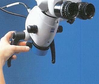
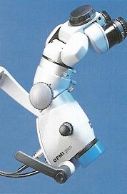
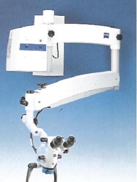
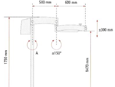
Of course dentists take care of their eyes. In this field there should not be a compromize. Therefore we choose the microscopes of Carl Zeiss. A brand (German), famous all over the world when things concern optical systems. On Zeiss microscopes all possible systems can be placed (modular) to react on your special demands. The dental Zeiss Operating Microscope is the only one with an integrated camera. Also design and the way to use the Zeiss microscope is absolute top class. Carl Zeiss microscopes belong to the best in the world. The famous endodontists from America subscribe this fully In the product catalogue stands the price of the microscope. Special offers are possible. If you want to have more information you can get it by mailing to:microscoop@endoplaza.com You will get an answer within one or two days. More information you can also find on www.beterlichtenzicht.nl The quality of the Operating MicroscopeA Microscope is an investment which one does once in a lifetime, therefore it is very important to make the right choice. You need the best optical systems. The stability of the arm of the microscope and the solidity of the mechanical parts should be perfect. Extra components, 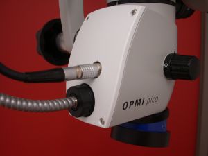 like a video camera (Zeiss is the only brand which integrates the camera in the microscope house) or inclineable binoculars 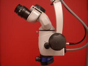 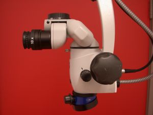 Can be realised simply. Therefore also a good support of the optical firm is important. A good microscope can be used during the full time practice of a dentist. At the end a microscope still is worth some money. Top class optical components are characterised by perfectly polished and coated lenses. There will be a brilliant and precise image reproduction. Lost of light is minimal. The importance of good mouth mirror 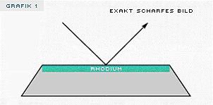 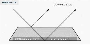 The technical data of the Microscope (steps of magnification, distance to the object, magnification, light goes in the same direction as the eyes of the operator, depth of field, field of view et cetera)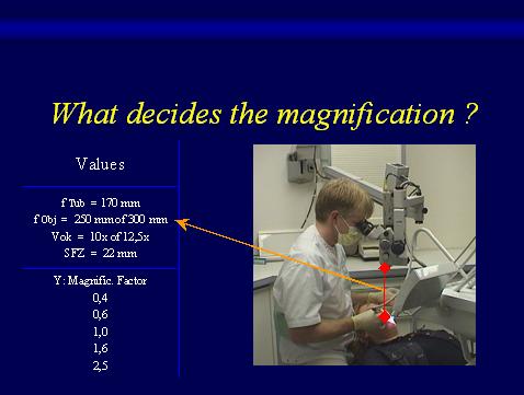 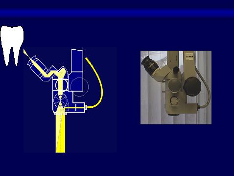 A microscope normally has 5 steps of magnification. (3,4,7,11 and 17) Factors which influence the magnification are:working distance (300mm or 250 mm lens), the binoculars (10 or 12,5) and the tubus. Besides the magnification, the light is very important. The coaxial light comes with the view of the operator in a corner of 4-6 degrees on the working field. In this way there is enough contrast and there is no trouble with shadow. 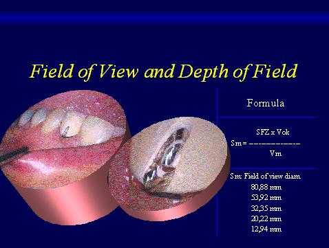 The components which make work more easyStandard the Opmi Pico is delivered with a 45 degrees inclined binocular. This means that you look in the microscope always in the same way. Inclineable binoculars give you the opportunity to vary in the direction you want to look. An extra 60 degrees extender gives you the opportunity to look with direct view to many parts of the mouth. The mostly used objective lenses are 250 or 300 millimeters (the working distance can vary depending on the length of the operator). A Beam-splitter is a possibility to divide the image in more directions (camera, video and extra binoculars. The Opmi Pico also can be delivered with a video which is integrated in the house of the microscope. In what parts of dentistry the Operating Microscope has benefits
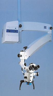
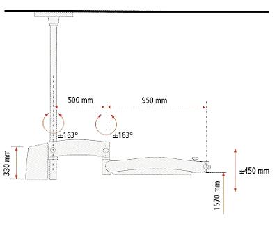
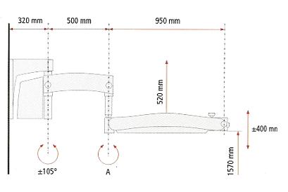
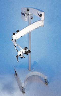
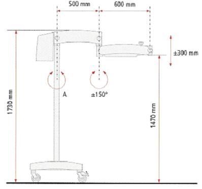
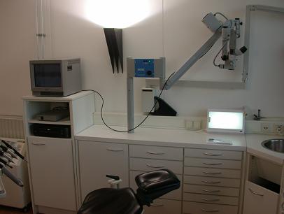
How to reduce or restore shoulder, neck and back stain (ergonomics)To sit in the right angle, many complaints of neck and shoulder could be prevented. Already existing complaints sometimes disappaer spontaneously. A dynamical working position is essential. One can realise this easily with a Microscope. The angle between neck and back should be no more than 25 degrees. 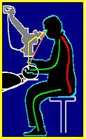 How to implementate the Operating Microscope in your practiceIt is important to think twice before buying a Microscope. Where sould the Microscope be placed? It should not interfere with the dentist or his assistance. In Hoogezand (The Netherlands) all 4 hereafter mentioned options are available. The docents of the course "The Operating Microscope in Dentistry" will advise you about your specific situation. The four possibilities to place the Microscope in your office.The possibilities are:
Documentation (digital) from treatmentsFotographs and video films can be used with the aid of an integrated camera. It is possible to put the documentation on DVD,VHS or in a digital (röntgen) system. Why are not the eyes tired after a whole day of working with the MicroscopeBecause your eyes do not have to accommodate nor to convergate, (Look to the power point presentations) How come that one can work faster and more predictableRoot canals are more easily to find. You can read the floor of the pulp chamber. You see what you do. The outline of a preparation is to see perfectly. The neighbours will not be prepared by accident.
|